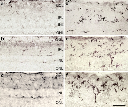
Fig. 1.
Distribution and morphology of OX42-immunoreactive microglia in cross-retinal sections. In normal retina (a), only processes without cell body showing OX42- immunoreactive can be detected. In the OH retina from the PBS feeding group (b), ramified microglia can be detected. In the OH retina of animals receiving different doses of the Wolfberry extract, 1 mg/kg (c), 10 mg/kg (d) and 100 mg/kg (e), there was an increase both in the number and immune intensity of OX42 microglia. In the 1000 mg/kg group (f), increased number of fully activated microglia was detected. They contained coarse and swollen perikarya that connected with thick processes. Scale bar is 50 μm

