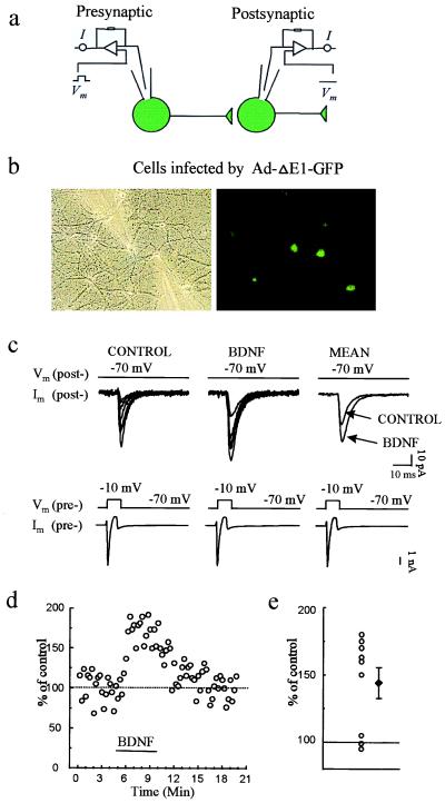
Figure 2.
Infection by the control Ad-ΔE1-GFP reporter virus allows BDNF-induced potentiation of evoked synaptic transmission. (a) Scheme of recording arrangement indicating that whole-cell recordings were obtained from two synaptically connected neurons, both expressing GFP. (b) Phase-contrast and fluorescence images of GFP-expressing cells during recordings. (c) Representative voltage-clamp currents in presynaptic (Lower) and postsynaptic (Upper) cells; the postsynaptic traces show EPSCs. Recordings are shown before (Control) and during exposure to BDNF (3 min after the start of BDNF perfusion). Traces labeled MEAN are averaged from eight consecutive EPSCs before or after BDNF application. The EPSC amplitude before and during BDNF application was 103.5 ± 19.1 and 147.2 ± 28.6 pA, respectively (mean ± SEM). (d) Plot of EPSC amplitude in an exemplar experiment before, during, and after BDNF application. (e) Percentage of potentiation (at 3 min after start of perfusion with BDNF) for each of the nine individual pairs studied (○). ♦, mean and SEM.

