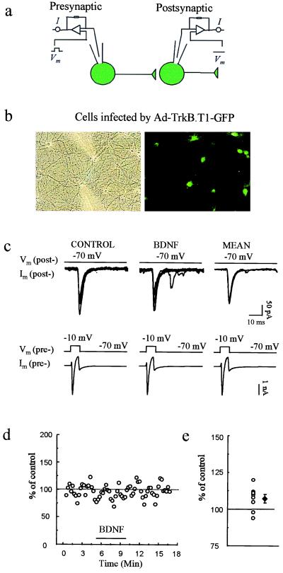
Figure 3.
Ad-TrkB.T1-GFP infection of both pre- and postsynaptic neurons prevents BDNF-induced potentiation of evoked synaptic transmission. (a) Scheme of recording arrangement indicating that whole-cell recordings were obtained from two synaptically connected neurons, both infected with Ad-TrkB.T1-GFP. (b) Phase-contrast and fluorescence images of GFP-expressing cells during recordings. (c) Representative individual and averaged pre- and postsynaptic traces, as in Fig. 2, showing EPSCs elicited before (CONTROL) and during BDNF application. The mean EPSC amplitude before and after BDNF application was 94.3 ± 21.9 and 98.1 ± 21.3 pA, respectively. (d) Plot of EPSC amplitude in an exemplar experiment before, during, and after BDNF application. (e) Percentage of potentiation (at 3 min after the start of perfusion with BDNF) for each of the eight pairs studied (○). ♦, mean and SEM.

