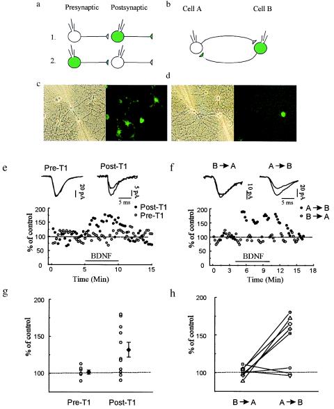
Figure 4.
Infection of presynaptic, but not postsynaptic, neurons with Ad-TrkB.T1-GFP prevents BDNF induced potentiation of evoked synaptic transmission. Whole-cell recordings were obtained from 26 pairs of synaptically connected neurons comprising one infected neuron (shown as green) and one noninfected neuron (shown as clear). (a) Scheme of recording arrangement for 18 of the pairs. In seven pairs, the infected neuron was presynaptic; in the other 11 pairs, the infected cell was postsynaptic. (b) Scheme of the recording arrangement for eight other pairs that were reciprocally connected; depolarizing pulses were applied alternatively in each cell, eliciting EPSCs that were measured in the other cell. (c and d) Phase-contrast and fluorescence images of exemplar fields during the recordings schematized in a and b, respectively. (e) Either the pre- (Pre-T1, ○) or postsynaptic (Post-T1, •) neuron was infected with Ad-TrkB.T1-GFP. When the infected neuron was postsynaptic BDNF potentiated the evoked response. Interleaved recordings in which the infected neuron was now presynaptic, revealed that BDNF failed to potentiate evoked transmission. The mean EPSC amplitude before and after BDNF application was 111.8 ± 17.6 and 152.2 ± 26.0 pA, respectively, for pairs with Post-T1. The mean EPSC amplitude before and after BDNF application was 99.9 ± 24.0 and 100.7 ± 24.4 pA, respectively, for pairs with Pre-T1 neurons. (g) Percentage of potentiation (at 3 min after the start of BDNF perfusion) for each pair (○). ♦, mean and SEM. (f) Plot of EPSC amplitude for an exemplar experiment before, during, and after BDNF application for the reciprocally connected pairs schematized in b. During BDNF application, transmission was potentiated when the infected neuron was postsynaptic (•, A → B), but not when the infected neuron was presynaptic, even though the tests were performed during interleaved 1-min intervals. The mean EPSC amplitude before and after BDNF application was 111.8 ± 17.6 and 152.2 ± 26.0 pA, respectively, for stimuli applied to the nonexpressing neuron and recorded postsynaptically in the T1-expressing neuron (A → B). The mean EPSC amplitude before and after BDNF application was 99.9 ± 24.0 and 100.7 ± 24.4 pA, respectively, for stimuli applied to the T-expressing neuron and recorded in the nonexpressing postsynaptic neuron (B → A). (h) Mean percentage of potentiation (at 3 min after start of BDNF perfusion) for each individual experiment with the reciprocally connected pairs.

