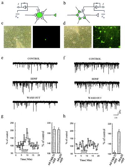
Figure 5.
Expression of Ad-TrkB.T1-GFP in presynaptic but not postsynaptic neurons inhibits BDNF-induced increases in mEPSC frequency. (a and b) Schemes of recording arrangement indicating that whole-cell recordings were obtained from single neurons that were either (a) infected with Ad-TrkB.T1-GFP and innervated by many uninfected neurons or (b) uninfected but innervated by many infected neurons. (c and d) bright-field and fluorescence images of GFP-expressing cells during recording schematized in a and b, respectively. (e) Representative sweeps containing mEPSCs before, during, and after BDNF application in an exemplar experiment schematized in a. (g) Plot of mEPSC frequency before, during, and after BDNF application in an experiment like that shown in e. The mean percentage of control is expressed relative to values obtained from uninfected cultures at 3 min after the start of BDNF perfusion. (f) Representative sweeps containing mEPSCs before, during, and after BDNF application in an exemplar experiment schematized in b. (h) Plot of mEPSC frequency before, during, and after BDNF application in an experiments like that in f. Mean percentage of control is expressed relative to values obtained from uninfected cultures at 3 min after the start of BDNF perfusion.

