
Figure 1. Assay for determination of Nuclear DNA-end binding activity.
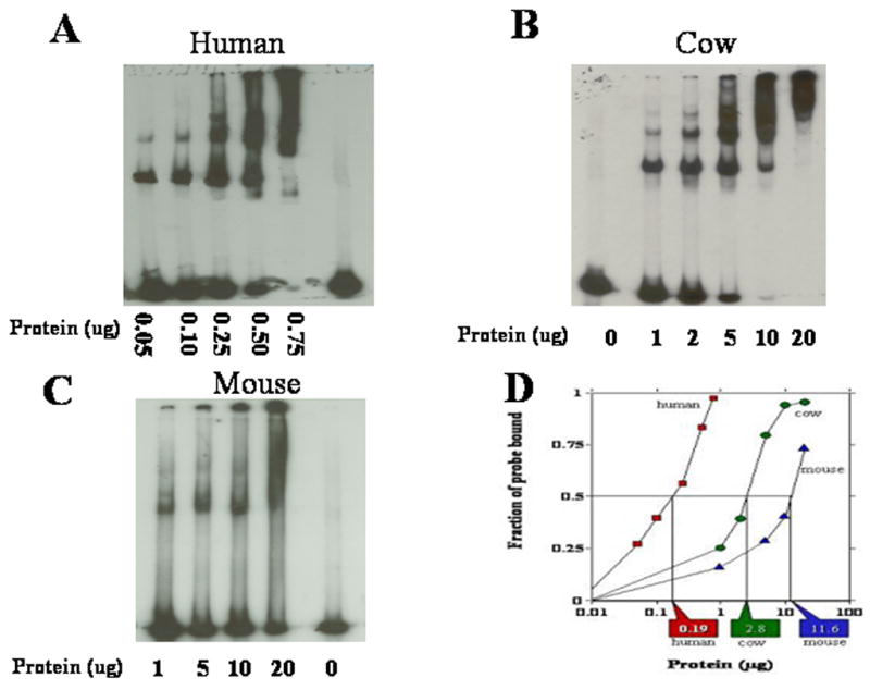
Typical mobility shift assays for the determination of DNA-end binding activity are shown for nuclear protein extracts isolated from human (A), mouse (B) and cow (C) fibroblasts. (D) Quantification of phosphorimager scans of DNA probe binding for each mobility shift. Vertical lines and rectangular callouts represent that amount of protein necessary to bind 50% of the 32P-labeled probe of linear DNA.