
Figure 6. Telomere length across mammalian species with different longevity.
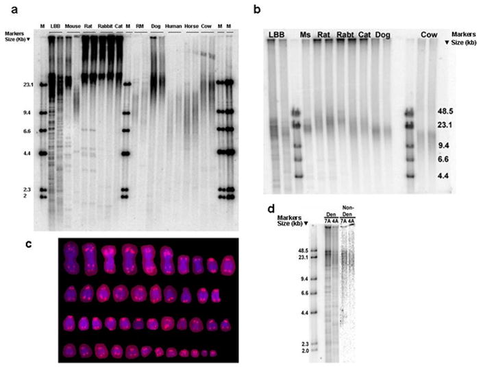
(a) Digested genomic DNA was resolved on a 0.5% agarose gel and probed with an end-labeled (CCCTTA)4 oligonucleotide. Species are ordered by increasing body mass (in grams). DNA marker lengths are shown in kilobases. The first line of the mouse is derived from a wild caught mouse from Pennsylvania, the second line is derived from a wild caught mouse from Idaho. RM = Rhesus monkey. M = DNA ladder. (b) Pulse field gel electrophoresis was used to resolve the telomeres that were too long to be measured in (a), above. Ms = Pennsylvania wild caught mouse, Rabt = rabbit. (c) Fluorescence in situ hybridization (FISH) with a Cy3-conjugated peptide nucleic acid probe (CCCTAA)3 showing that little brown bat telomeres contain internal telomeric repeats. (d) Little brown bat (LBB) telomeres were probed under denaturing conditions (left) or non-denaturing condition (right), only the telomere signals that were detected under non-denaturing conditions were used to estimate mean telomere length. For the majority of the lines the population doublings of the culture were 25 and cells had not immortalized. Mouse and rat cells had spontaneously immortalized.