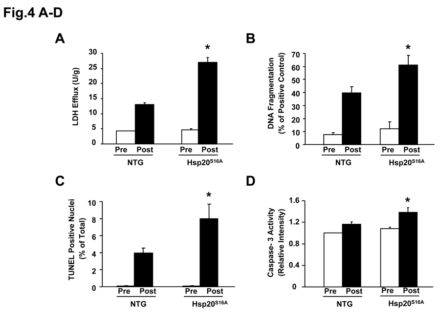
Figure 4.
Hsp20S16A overexpression increased ischemia/reperfusion-induced necrosis and apoptosis. At basal level, there is no significant differences in LDH release (A), DNA fragmentation (B), TUNEL positive nuclei (C) and caspase-3 activity (D), between Hsp20S16A and non-transgenic hearts (non-TGs: n=6, Hsp20S16A: n=6, P>0.05). Hsp20S16A hearts, subjected to no-flow ischemia followed by reperfusion, exhibited significantly increased total LDH release (A), DNA fragmentation (B), TUNEL-positive nuclei (C) and caspase-3 activity (D), compared to non-TGs (non-TGs: n=6, Hsp20S16A: n=6, *: P <0.01 vs. non-TG).

