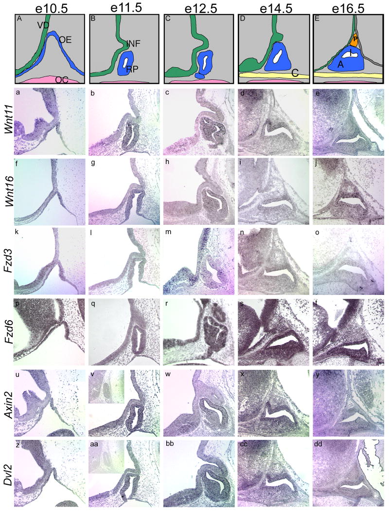
Figure 7. Spatial expression patterns of Wnt pathway members in the developing pituitary and ventral diencephalon.
Formation of Rathke's pouch is illustrated from e10.5 to e16.5 (A-E). At e10.5, the oral ectoderm begins to invaginate and pinches off around e11.5 to form Rathke's pouch. Meanwhile, the ventral diencephalon evaginates to form the infundibulum. From e12.5 to e14.5, the anterior lobe begins to form as cells surrounding the lumen rapidly divide and migrate out of the pouch. By e16.5, the three distinct lobes of the developing pituitary are evident, with the posterior lobe arising from the infundibulum. In situ hybridizations were performed on wild-type sagittal sections from e10.5 to e16.5 to determine temporal and spatial patterns of expression. Slides are oriented with dorsal to the top and rostral to the left. Inset panels (v, aa) show sense slides for negative controls. VD, ventral diencephalon; OE, oral ectoderm; OC, oral cavity; INF, infundibulum; RP, Rathke's pouch; C, cartilage plate of hard palate; P, developing posterior lobe; I, developing intermediate lobe; A, developing anterior lobe.

