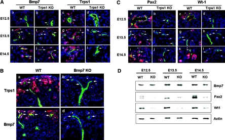
Figure 4.
Comparison of Bmp-7, Trps1, Pax2, and Wt1 expression during early renal development. (A) Double immunofluorescence staining of kidneys obtained at E12.5 (a through d), E13.5 (e through h), and E14.5 (i through l) from WT mice (a, e, i, c, g, and k) and Trps1 KO mice (b, f, j, d, h, and l) with antibodies for Bmp7 (red) and cytokeratin (green) (a, b, e, f, i, and j) or Trps1 (red) and cytokeratin (green) (e, d, g, h, k, and l). (B) Double immnofluorescent staining of kidneys obtained at E14.5 from WT mice (a and c) and Bmp7 null KO mice (b and d) with antibodies for Trps1(red) and cytokeratin (green) (a and b) or antibodies for Bmp7 (red) and cytokeratin (green) (c and d). (C) Double immunofluorescence staining of kidneys obtained at E12.5 (a through d), E13.5 (e through h), and E14.5 (i through l) from WT mice (a, e, i, c, g, and k) and Trps1 KO mice (b, f, j, d, h, and l) with antibodies for Pax2 (red) and cytokeratin (green) (a, b, e, f, i, and j) or Wt1 (red) and cytokeratin (green) (e, d, g, h, k, and l). The ureteric buds (arrows) are positive for cytokeratin. Arrowheads and asterisks indicate cap mesenchyme and renal vesicles, respectively. Nuclei are stained with DAPI. (D) Western blot analysis of Bmp7, Pax2, and Wt1 in protein extracts from E12.5, E13.5, and E14.5 kidneys of WT mice and Trps1 KO mice.

