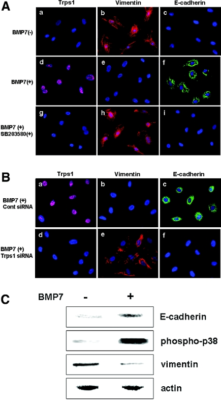
Figure 5.
RMMs are transformed into epithelial cells through Bmp7/p38 MAPK/Trps1 signaling. (A) Expression of Trps1 (pink; a, d, and g), vimentin (red; b, e, and h), and E-cadherin (green; c, f, and j) by metanephric mesenchymal cells without treatment (a through c), with Bmp7 treatment (d through f), and with treatment by Bmp7 plus SB203580 (g through i). Nuclei were stained with DAPI (blue). (B) Expression of Trps1 (pink; a and d), vimentin (red; b and e), and E-cadherin (green; c and f) by metanephric mesenchymal cells with or without treatment by Bmp7 and control siRNA (a through c), or with treatment by Bmp7 and Trps1 siRNA (d through f). Nuclei were stained with DAPI (blue). (C) Western blot analysis of E-cadherin, phosphorylated p38 MAPK, and vimentin in extracts of cultured primary metanephric mesenchymal cells from rat E13.5 kidneys.

