Figure 1.
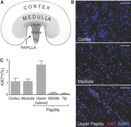
Cellular proliferation in adult kidney is shown. (A) Kidney regions were examined for cellular proliferation. (B) Ki67-positive cells in representative sections of kidney cortex, medulla, and upper papilla are shown. Cell nuclei stained with DAPI (blue) and Ki67 stained with rhodamine (red) are shown. Bars = 50 μm. (C) Fraction of Ki67-positive cells is shown. Cortex and medulla had a similar number of Ki67-positive cells, but the lateral side of the upper papilla had significantly more Ki67-positive cells than both the cortex and the medulla as well as other parts of the papilla (P < 0.01). The tip and middle part of the papilla had significantly fewer Ki67-positive cells than all other areas of the kidney (P < 0.01).

