FIGURE 1.
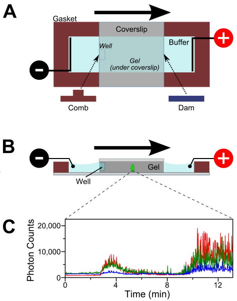
Schematic of a mini-gel assembly for in-gel ALEX. (A) Top view of the gel apparatus. Polyacrylamide (PA) is polymerized between two glass coverslips spaced by a 2-mm silicone gasket. The electric field is supplied through two platinum electrodes immersed in the running buffer. Sample is loaded in the well (formed by leaving a small comb during polymerization process). Negatively charged biomolecules (e.g., DNA) migrate towards the positive electrode (migration direction denoted by the bold arrow). The migration rate is determined by the charge and size of the biomolecules.
(B) Cross-section of the mini-gel assembly. The confocal volume (green ellipse; not to scale) is positioned halfway in the gel along the direction of electrophoresis and ≈30 μm above the top surface of the bottom coverslip.
(C) Fluorescence intensity traces from a stationary confocal spot show the appearance of fluorescence bands as labeled molecules undergo electrophoresis through the confocal volume. Here, an RNA polymerase (RNAP) open complex mixture was separated in 5% PA and observed under ALEX. The time traces were binned with 1-s resolution. Green: fluorescence in the donor-emission channel due to donor excitation (FDex,Dem); blue: fluorescence in the acceptor-emission channel due to donor excitation (FDex,Aem); red: fluorescence in the acceptor channel due to acceptor excitation (FAex,Aem). The burst centered at ≈3.5 min corresponds to the free doubly labeled DNA; the burst starting at ≈10 min is the (RNAP)-DNA open complex.

