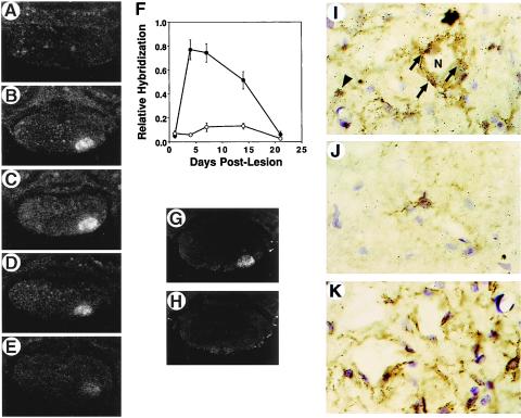
Figure 4.
ISH analysis of CX3CR1 expression in the rat facial nucleus after motor neuron axotomy. ISH was performed on rat brainstem sections from animals killed at 1 (A), 4 (B), 7 (C), 14 (D), and 21 (E) days after nerve transection by using [35S]UTP-labeled riboprobe. F depicts a quantitative analysis of the hybridization signal on the operated (•) and contralateral (○) sides. Results are presented as the mean ± SEM of data derived from at least three sections from three different animals. The hybridization signal was specific for CX3CR1 because no increase in signal was observed by using a sense riboprobe (H) when compared with a corresponding anti-sense riboprobe hybridized-section (G). Hybridization signals (silver grains) from anti-sense [33P]UTP-labeled riboprobe hybridized-sections are found principally over lectin (GSA-I-B4)-staining microglia in the neuropil of the unoperated facial motor nucleus (J) or lesioned facial motor nucleus (arrowhead in I) whereas several CX3CR1 mRNA-containing microglial cells are found perineuronally (arrows in I) surrounding axotomized motor neurons (N, motor neuron cell body). (K) Sense [33P]UTP-labeled riboprobe-hybridized section showing only a few scattered silver grains.

