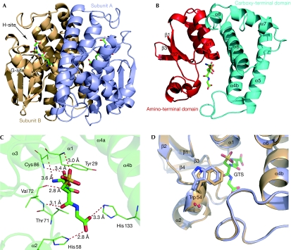
Figure 1.
Overall structure of Gtt2. (A) Schematic representation of the Gtt2 homodimer. GTS molecules are shown as stick models and coloured according to their atom type (C, green; S, yellow; O, red). The G-site and H-site of subunit B are indicated by arrows. (B) The GTS-bound Gtt2 monomer, with the two domains in cyan and red. (C) Hydrogen bonds between GTS and Gtt2. (D) Superposition of apo-bound (light blue) and GTS-bound Gtt2 (light orange) showing the different position of the loop after helix-α2 upon GTS binding. Cys, cysteine; G-site, GSH-binding site; GTS, glutathione sulphonate; H-site, hydrophobic substrate binding site; His, histine; Thr, threonine; Trp, tryptophan; Tyr, tyrosine; Val, valine.

