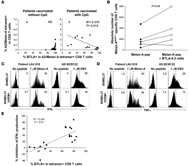
Figure 6. Functional inhibition of CD8+ T cells depending on BTLA expression and HVEM triggering.
(A) Direct ex vivo analysis of percentage of A2/Melan-AMART-1 tetramer+ CD8+ T cells among PBMCs, in relation to their BTLA expression, after vaccination without and with CpG. (B) In vitro expansion of Melan-AMART-1 tetramer+ T cells from healthy donors, after 10 days of stimulation with peptide-loaded DCs, in the absence versus presence of blocking mAb BTLA-8.2. (C and D) IFN-γ (C) and TNF-α (D) production by BTLA+ antigen-specific CD8+ T cells after 4 hours of peptide stimulation of PBMCs assessed ex vivo, i.e., without prior in vitro cultivation. Representative histograms of PBMCs from patient LAU 618 (after 2 vaccinations, with CpG) and from healthy donor BCB122 stimulated by SKMEL37 cells expressing HVEM or not, loaded with Melan-AMART-1 or EBV peptides, respectively. Histograms are gated on tetramer+ T cells. Percentages of cytokine-positive cells are indicated. (E) Significant correlation between percentages of BTLA+ tetramer+ T cells and percentages of IFN-γ production inhibition (i.e., reduction when stimulated with HVEM+ SKMEL37 cells as compared with HVEM– SKMEL37 cells). Each dot represents a single experiment with Melan-AMART-1– or EBV-specific T cells.

