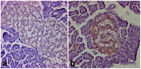
Figure 4. Comparison of OX1R-immunoreactivity in the endocrine pancreas of Wistar and GK rats.
Light micrographs showing OX1R-immunoreactive cells in the pancreatic islet of normal Wistar (a) and Goto Kakizaki (b) rats. Note that the islet cells of Goto Kakizaki stains more intensely for OX1R compared to Wistar. Magnification: ×200.

