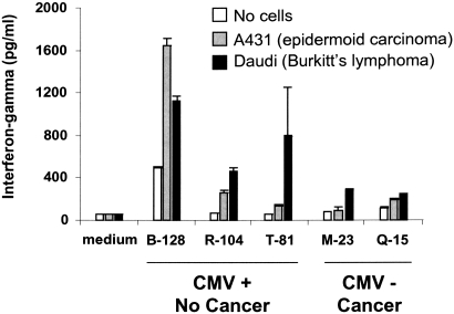
Figure 3.
In vitro antitumor reactivity of CMV-induced Vδ2neg γδ T cells. γδ T cells from either patients who developed CMV-infection and did not display cancer (CMV+ cancer-free) or patients who did not develop any CMV infection but displayed a cancer (CMV-free cancer+) were sorted from PBMCs. γδ T cells were then expanded in culture RPMI medium supplemented with 10% human serum, 1000 U/ml rIL-2, 15 ng/ml rIL-15, and irradiated autologous PBMCs. After 1 mo, those γδ T cell lines were incubated with the A431 (epidermoid carcinoma) and the Daudi (Burkitt's lymphoma) cell lines for 24 h in the presence of rIL-12 and rInterferon-α. IFN-γ released into the supernatant was quantified by ELISA.

