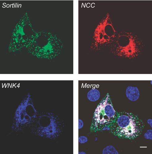
Figure 8.
NCC co-localizes with sortilin and WNK4. Cos-7 cells were co-transfected with myc-WNK4 WT, GFP-sortilin WT, and HA-NCC WT. Forty-eight hours after transfection, immunostaining and confocal microscopy were performed. The distribution pattern for sortilin WT in green seems to be similar to sortilin WT as described in Figure 5A, and NCC expression pattern in red seems to be similar to the NCC pattern as described in Figure 5B. WNK4 in blue is expressed in the perinuclear and cytoplasmic regions. The merged picture shows the co-localization of WNK4, sortilin, and NCC in the perinuclear region in white, indicating that WNK4, sortilin, and NCC might be associated together.

