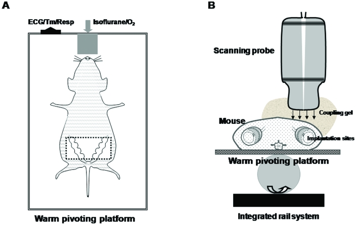
Figure 1.
Positioning and ultrasound imaging of an anesthetized, dorsally recumbent pregnant mouse (A) as an arial view and (B) in transverse cross-section. Studies can be performed on mice at different gestational stages. All hair was removed from the ventral abdomen after the mouse was anesthetized with approximately 2.0% (1.5% to 2.5%) isoflurane by means of an oxygen mask. The mouse then was placed on the platform and held in position with surgical tape. A thick layer of warm water-based coupling gel was applied over the skin of the area to be imaged. A 40-MHz transducer probe was applied to the skin to collect images. The transabdomenal area (boxed region in A) is suitable for detection of the pregnant uterus. To acquire optimal images, the scanning probe needs to carefully be adjusted to the natural orientation of each implantation site. All waveforms are saved for later offline analysis. Maternal heart and respiration rates were monitored by using an autom ated system. Body temperature was maintained as 36 to 37 °C by the warmed platform, which is supported by an integrated rail system. Figures are not to scale.

