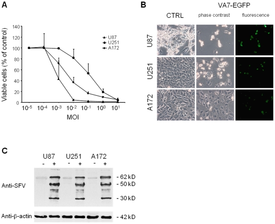
Figure 1. VA7-EGFP kills glioma cells in vitro.
(A) Dose-dependent killing by VA7-EGFP of U87, U251 and A172 glioma cells in culture. Glioma cells were seeded in a 96-well plate and VA7-EGFP at MOI ranging from 10−5 to 101 was added 16 hours later. Cell viability was assessed 96 hours later using calcein AM as indicator of cell membrane integrity. The mean values and standard deviations are shown. (B) Photograph of the cells at 96 hours post infection with VA7-EGFP at MOI 0.1. Fluorescence channel for VA7-EGFP shows GFP-positive cells indicative of active virus replication. (C) Western blot of VA7 structural protein expression in untreated and VA7-EGFP -infected cells 18 hours post infection. Equal number of cells were infected at MOI 1 and collected for Western analysis 18 hours later. In all cell lines the 30 kDa capsid, the E1 and E2 50 kDa proteins and the E2 precursor p62 were abundantly expressed.

