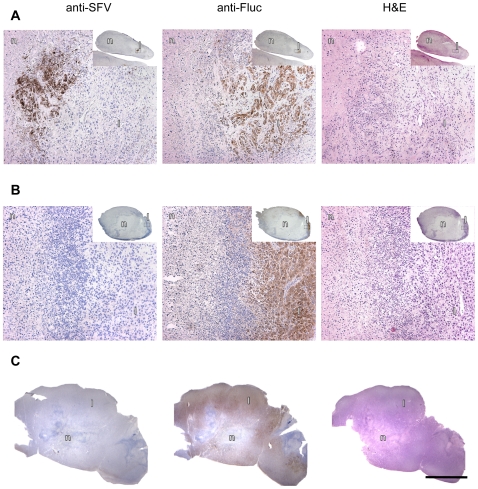
Figure 3. Histological analysis of subcutaneous U87Fluc tumor xenografts.
Anti-SFV immunostaining, anti-luciferase staining and H&E staining, respectively, of parallel sections of residual tumor tissue in a mouse 7 days after injection with a single intravenous dose of 1×10⁶ PFU of VA7-EGFP (A) or 21 days after the third injection of VA7-EGFP (B) (original magnification ×200, insert ×10). (C) Similar staining of a xenograft from a PBS-treated tumor-bearing mouse. n = necrotic tissue, l = live cells. Bar = 5 mm.

