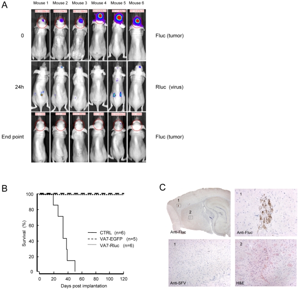
Figure 5. A single intravenous injection of VA7-Rluc has potent antiglioma effect in nude mice bearing orthotopic U87Fluc tumors.
(A) Orthotopic U87Fluc xenografts were established in Balb/c nude mice by stereotactic injections of the tumor cells. When the firefly bioluminescence from the tumor xenografts reached appropriate levels the animals were given an intravenous injection of 1×10⁶ PFU of VA7-Rluc. After 24 hours, the animals were injected intravenously with ViviRen substrate and the light produced by the VA7Rluc vector was monitored by a cooled CCD camera. Transient virus replication (Rluc) could be detected in the brain, co-localizing with the tumor (Fluc), as well as in the periphery. Even tumors close to endpoint were fully eradicated. Virus signals can be compared with each other but not with tumor signal due to different intensity scale. (B) Long-term survival of mice in Figure 4 and Figure 5A. None of the animals treated intravenously with either VA7-EGFP or VA7-Rluc reached the end point bioluminescence value. The median survival of PBS-treated (CTRL) mice was 36 days (95% CL = 26 days to 42 days) and all differences to this group were highly significant (log-rank test, P<0.0001). (C) Sagittal section of the brain of mouse 5 stained against Fluc (top left). This mouse was given a single intravenous dose of 1×10⁶ PFU of VA7-Rluc and was the only one in the group of intravenously -treated mice that produced an intracranial Fluc IVIS signal at the final measurement of the experiment. Top right, 20× magnification of box 1 showing a Fluc-positive cell cluster. Bottom left, 10× magnification of box 1 showing anti-SFV immunostaining of parallel brain section from the same mouse. No SFV immunoreactivity could be detected anywhere in the brain of this or any other i.v. treated mouse. Bottom right, 10× magnification of the region in box 2 from a parallel section stained with H&E. The increased cellular density surrounding the blood vessel in the picture is indicative of regenerative activity. This region was possibly the site of the original tumor before destruction by the VA7-Rluc virus.

