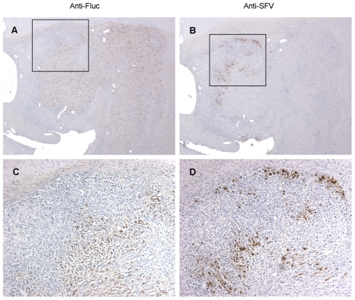
Figure 6. Immunohistochemical analysis of VA7 replication in intracranial U87Fluc xenografts.
U87Fluc cells were implanted in the striatum Balb/c nude mice and tumor growth was monitored by magnetic resonance imaging. When tumors were visible mice were injected with 1×10⁶ pfu of VA7-EGFP and sacrificed three days later. Brains were removed and paraffin sections prepared. (A–D) Representative images of U87Fluc xenograft immunostained against Fluc (A,C) and SFV antigen (B,D). (A,B) magnification, ×27; (B,D) 3.7× magnification of boxed areas in (A,B).

