Figure 1.
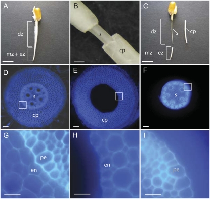
A to C, A 2.5-d-old seedling root of the inbred line B73 before (A) and after (C) separation of stele and cortical parenchyma in the differentiation zone of the root. A, The differentiation zone (dz) is marked by the presence of root hairs, while the root tip contains the meristematic zone (mz) and the elongation zone (ez). B, Close-up after cutting the cortical parenchyma close to the coleorhiza and removing the cortical parenchyma (cp) from the stele tissue (s). C, Differentiation zone of the same seedling as in A after removing the root tip (mz + ez) and mechanical separation of the cortical parenchyma (cp). Subsequently, these tissues were used for protein and hormone analyses. D to I, Toluidine blue-stained free hand transverse sections of the differentiation zone of 2.5-d-old B73 primary roots before (D and G) and after (E, F, H, and I) manual separation. D, Transverse whole root transverse section before manual separation. E, Transverse section of the cortical parenchyma after separation (compare with cp in B and C). F, Transverse section of the stele after separation (compare with s in B and C). G, Close-up of the region marked by a white box in D, indicating the junction of pericycle (pe) and endodermis (en) cell layers. H, Close-up of the region marked by a white box in E. Note the intact endodermis (en) cell layer after tissue separation. I, Close-up of the region marked by a white box in F. Note the intact pericycle cell layer (pe) after tissue separation. Bars = 1 cm (A and C), 500 μm (B), 100 μm (D–F), 50 μm (G–I). [See online article for color version of this figure.]

