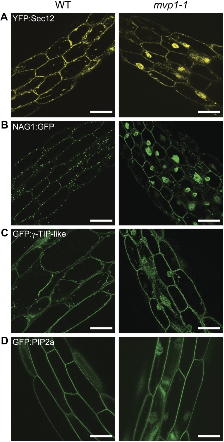
Figure 2.
The mvp1-1 mutation disrupts protein targeting to the ER, Golgi, vacuole, and plasma membrane. Confocal images of 7-d-old hypocotyl tissues show that the mvp1-1 mutation leads to partial aggregation of endomembrane fusion proteins. A, YFP:Sec12 in the ER. B, Golgi fusion NAG1:GFP. C, Tonoplast protein GFP:γ-TIP-like. D, Plasma membrane fusion GFP:PIP2a. WT, Wild type. Bars = 50 μm.

