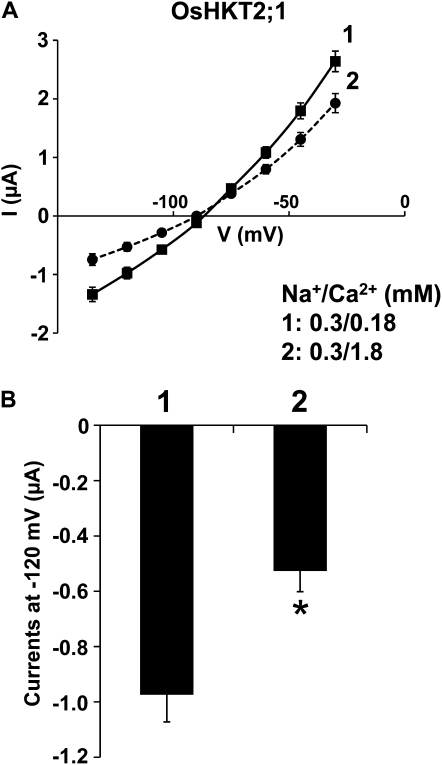
Figure 7.
Increasing the external Ca2+ concentration reduces OsHKT2;1-mediated Na+ currents in Xenopus oocytes. A, Average current-voltage curves from OsHKT2;1-expressing oocytes bathed in a 0.3 mm Na+ solution containing either 0.18 mm Ca2+ (solid line, no. 1) or 1.8 mm Ca2+ (dashed line, no. 2). Currents were measured using a step command with 15-mV decrements (see “Materials and Methods”). Error bars represent se (n = 13–15) from two independent experiments. B, Current amplitudes recorded at −120 mV, derived from OsHKT2;1-expressing oocytes in the presence of 0.18 mm Ca2+ (no. 1; n = 15) or 1.8 mm Ca2+ (no. 2; n = 13), which were extracted from the data sets presented in A. Error bars represent se (* P < 0.01, 0.18 mm Ca2+ versus 1.8 mm Ca2+).

