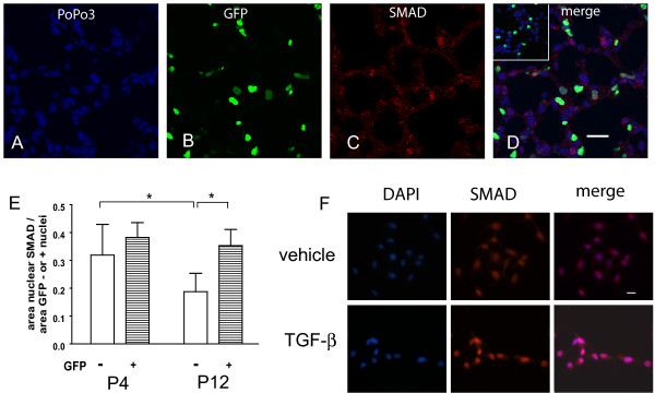
Figure 10.
PDGF-Rα expression does not preclude SMAD-dependent TGFβ signaling in the lung. Lung tissue from PDGF-Rα-GFP mice aged P4 and P12 was stained for SMAD 2/3 followed by Alexa Fluor-568-conjugated secondary antibody (red, C) and PoPo 3 iodide nuclear counterstain (blue, A). The area of nuclear SMAD 2/3 was determined from merged images (D) in the nuclei cells that either express PDGF-Rα (yellow- red SMAD merged with green GFP) or do not express PDGF-Rα (magenta - red SMAD merged with blue pseudocolored PoPo3) cells. The inset in D (secondary antibody only) shows minimal levels of Alexa Fluor-568 staining in the absence of the primary antibody. Scale bars are 20 μm. Results are summarized in the column graph (E). The error bars represent standard deviations from the averages for each group. *, P < 0.001. To verify that nuclear translocation was TGFβ dependent, Mlg cells were treated with either vehicle or 0.125 ng/ml TGFβ (F) for 30 min and stained for SMAD 2/3 followed by 4', 6-diamidino-2-phenylindole (DAPI) nuclear counterstain. More SMAD 2/3 localized to the nuclei of TGFβ-treated cells.

