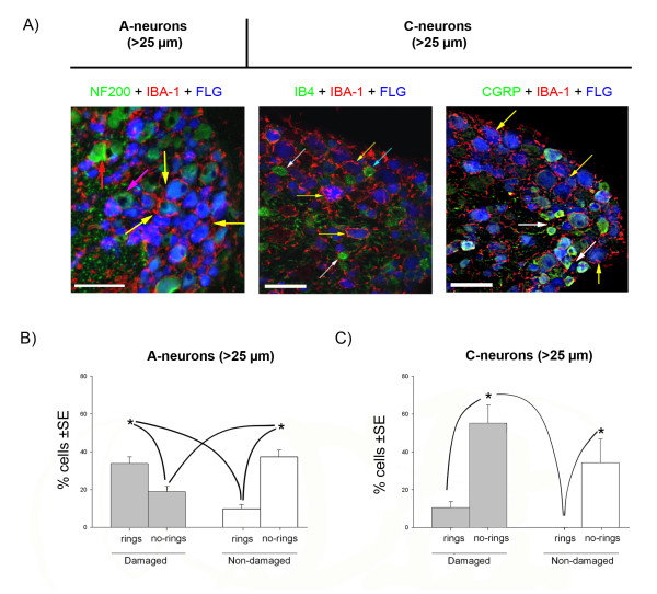
Figure 5.
Analysis of the macrophage invasion of adult dorsal root ganglia 7 days after spared nerve injury. Fluorogold (FLG, blue) was used as a retrograde tracer injected at the site of nerve injury, to label damaged neurons. A) Macrophages (IBA-1 positive, in red) surround large A-neurons (> 25 μm and NF200 +ve, in green) and are distributed as a 'ring-like' structures. The macrophages have large cell bodies and processes directed toward the damaged neurons (yellow arrows) and undamaged neurons (red arrow). The left panel shows that these 'ring-like' structures surround the many of Fluorogold labelled A-neurons (yellow arrows). Some non-Fluorogold labelled (undamaged) A-neurons are also surrounded by macrophages (red arrow) but others are not (pink arrow). The small diameter (< 25 μm) C-neurons (middle panel: non peptidergic, IB4 +ve, or right panel: peptidergic, CGRP +ve; both in green) do not generally display ring-like displays of macrophages (white arrows) although there are some exceptions (blue arrow). Scale bar: 100 μm. B) Graph showing the relative proportion of damaged (Fluorogold +ve) or undamaged A-neurons (> 25 μm) with macrophage rings (n = 559 A-neurons from 3 animals). C) Graph showing the relative proportion of damaged (Fluorogold +ve) or undamaged in C-neurons (< 25 μm) (n = 284 C-neurons, from 3 animals). Statistical significance is indicated by * (ANOVA p < 0.001, SNK p < 0.05, power>80%). Grey boxes indicate damaged neurons; white boxes indicate non-damaged neurons. Lines indicate the statistical differences between the several experimental groups. SE: standard error.

