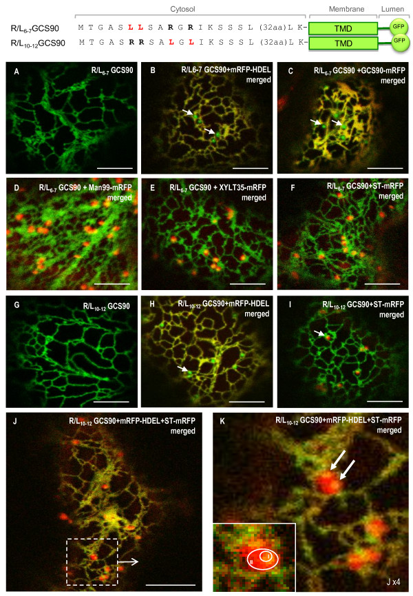
Figure 5.
Punctate structures do not accumulate ER resident proteins and are distinct from Golgi stacks. When arginine residues are mutated by pairs, R/L6-7GCS90 (A-I) and R/L10-12GCS90 (G-K) are located in the ER (A, G). Co-expression with soluble ER marker mRFP-HDEL (B, H) or membrane (C) ER marker GCS90-mRFP reveals those markers are excluded from the punctate structures that appear in green (arrows). Punctate structures are closely associated to Golgi stacks labelled with the cis-Golgi marker Man99-mRFP (D), the medial Golgi marker XYLT35-mRFP (E) or trans-Golgi marker ST-mRFP (F, I). When the constructs highlighting punctate structures are co-expressed together with the ER marker mRFP-HDEL and the Golgi marker ST-mRFP, the ER and the punctate structures appear in yellow (J). When zooming, micrograph suggests punctate structures can be closed to the ER (K, top and bottom arrows). Zone I corresponds to the co-localization area between a punctate structure and a Golgi whereas zone II corresponds to the Golgi only (K, insert). Arrows indicate the punctate structures.

