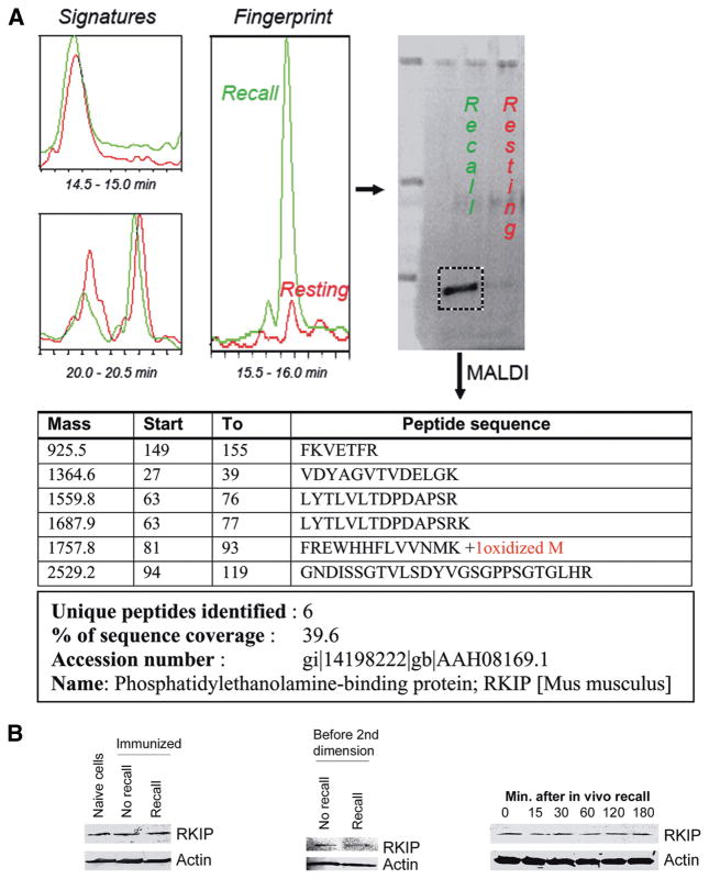
FIGURE 3.
Identification of RKIP as a fingerprint in recall cells. A, One second dimension fraction from the recall sample and its corresponding equivalent from the no recall sample were lyophilized and resolved by 4–15% SDS-PAGE. A 22 kDa band, detected by a protein-specific fluorescent dye was cut, digested by trypsin, sequenced by MALDI-TOF. Peptide sequences were searched against using the NCBInr database version 20060804 using the Proteometrics Software Suite and the Profound Search Algorithm. B, RKIP expression was assessed by immunobloting of the original sample (left), the cell lysate immediately before loading on the PF 2D (middle), and the kinetics of an in vivo recall response (right).

