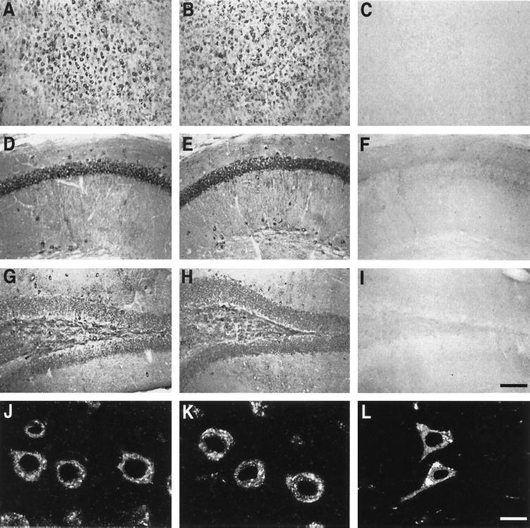
Figure 1.
Immunocytochemical detection of human apoE in neurons of NSE-apoE3 and NSE-apoE4 mice and a human with AD. (A–I) Immunoperoxidase staining for human apoE revealed widespread neuronal labeling, including neocortex (A–C), hippocampal CA1 region (D–F), and dentate gyrus (G–I), in NSE-apoE3 (A, D, and G) and NSE-apoE4 (B, E, and H) mice. No apoE labeling was seen in corresponding brain regions of a knockout control lacking NSE-apoE transgenes (C, F, and I). (J–L) Immunofluorescence staining for human apoE strongly labeled neocortical neurons in NSE-apoE3 (J) and NSE-apoE4 (H) mice, and in a human AD case (APOE ɛ3/ɛ4) (L). (Bars: A–I, 160 μm; J–L, 10 μm.)

