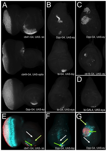
Figure 3. Ectopic eyes develop in a narrow range of cells within developing epithelial tissues.
(A-D) Confocal images of third instar eye, antenna, leg, wing and haltere imaginal discs. These are representative images from the screen demonstrating that ectopic eye formation is limited to sub-populations of cells within each epithelium. Photoreceptor cells are marked by the presence of ELAV. A = eye antennal disc, B = wing disc, C = leg disc, D = haltere. Anterior is to the right, dorsal is at the top.

