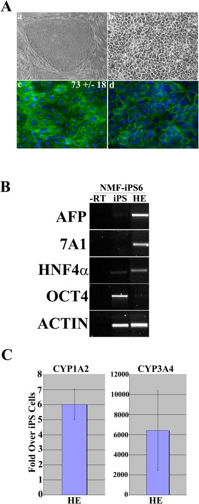
Figure 1. Derivation of hepatic endoderm from iPSCs.
(Aa) Phase contrast microscropy demonstrating the typical iPSC colony morphology and (Ab) iPSC-derived hepatic endoderm (HE) following 14 days in the differentiation procedure (Magnification x20). (Ac) iPSC-derived HE stains positive for albumin at day 14 in the differentiation procedure. The numbers represent the efficiency of the procedure +/− SE. (Ad) iPSC-derived HE stains positive for E-Cadherin at day 14 in the differentiation procedure (B) RT-PCR gene expression of iPSCs and iPSC-derived hepatic endoderm. iPSC-derived hepatic endoderm on (lane 1) express the markers AFP, CYP7A1 and HNF4 alpha, but not OCT-4. iPSCs strongly express OCT4. Potentially due to spontaneous differentiation found in iPSC culture. PCR reactions were controlled using a –RT control (lane 4) and details of primers and cycles can be found in the supplementary methods section (C) CYP1A2 and CYP3A4.

