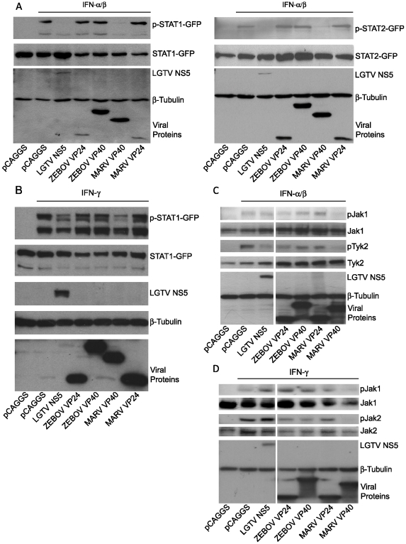
Figure 4. MARV VP40 inhibits IFN-induced STAT and Jak phosphorylation.
(A) STAT1 or STAT2 (1 µg) fused to a C-terminal GFP was co-expressed in Huh-7 cells with 2 µg of the indicated expression plasmids, treated with 1000 IU/ml of universal IFNα/β for 30 min. Cells were lysed and assayed by western blot for tyrosine phosphorylated STAT1 (p-STAT1-GFP), STAT2 (p-STAT2-GFP), as well as for total expression levels of over-expressed proteins. (B) Huh-7 cells were transfected with 2 µg of the indicated expression plasmids and 1 µg of a plasmid expressing STAT1 fused to GFP at the C-terminus (STAT1-GFP). 24h p.t., cells were treated with 1000 IU/ml of IFNγ for 30 min and lysed. Lysates were analyzed by western blot for phosphorylation of STAT1 and total levels of STAT1 as well as for expression of the tagged proteins. (C and D) 293T cells were transfected with 2 µg of the indicated expression plasmids, treated with 1000 IU/ml of IFNα/β (C) or IFNγ (D) for 30 min, lysed and subjected to western blot analysis for detection of phosphorylated and total Jak1 (C and D), Tyk2 (C) or Jak2 (D). Note that the cuts in the films excised samples irrelevant to this study. All the presented data for a given protein is from the same gel and the same exposure.

