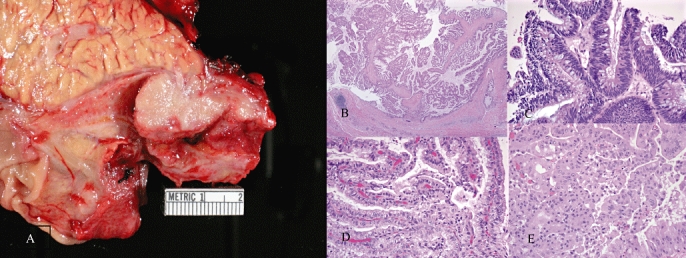
Figure 2.

Photograph A shows a distal common bile duct intraductal papillary mucinous neoplasm (IPMN) with characteristic gross polypoid features. Photographs B–E show haematoxylin and eosin stains of different biliary tract (BT)-IPMNs with papillary cytoarchitecture within the benign portion (b), intestinal epithelium (c), pancreatobiliary epithelium (d) and oncocytic epithelium (e)
