
Fig. 6.
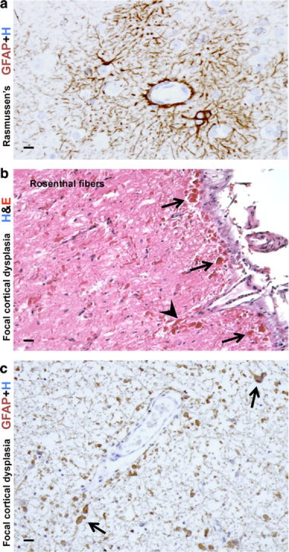
Reactive astrogliosis in two disorders with seizures. a High magnification image of cerebral cortex from an individual with Rasmussen encephalitis (RE). Immunohistochemical staining for GFAP shows moderate reactive astrogliosis that is especially prominent around small blood vessels. Note the intensely stained and hypertrophic astrocytic foot processes extending to and lining the adventitia of microvessels. b, c High magnification images of a resection specimen from an individual with severe seizures associated with severe focal cortical dysplasia stained for H&E (b) or GFAP (c). b In this unique case, abundant Rosenthal fibers were found throughout the resection specimen, especially in the subpial regions (arrows) and around blood vessels (arrowheads). The density of the Rosenthal fibers suggested the diagnosis of ‘focal’ Alexander’s disease. However, testing of the GFAP gene revealed no mutations (for details, see [111]). c Comparable section to b showing abundant GFAP immunoreactive astrocytes and associated Rosenthal fibers (arrows). Scale bars a 10 μm, b 20 μm, c 15 μm