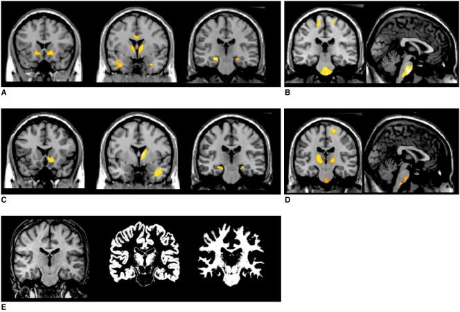
Fig. 3.
White matter concentration changes in mesial temporal lobe epilepsy patients. White matter concentrations reduced in internal capsules, temporal stem, and parahippocampal regions in left (A) and right (C) mesial temporal lobe epilepsy patients, whereas white matter concentrations increased in precentral gyri, pons, and internal capsules in left (B) and right (D) mesial temporal lobe epilepsy patients. Pons (arrow) was partitioned into white matter image using SPM2 segmentation (E). These findings were significant at uncorrected p < 0.001.

