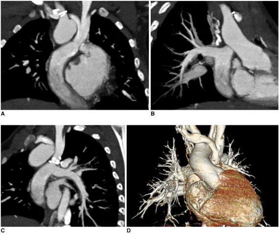
Fig. 12.
Non-ECG-synchronized multiplanar reformatted (A-C) and volume-rendered (D) CT images clearly show patent Fontan pathway and pulmonary vessels in 5-year-old boy with functional single ventricle. Simultaneous intravenous injection of 50% diluted contrast agent through arm and leg veins was used to obtain homogeneously high enhancement of cardiovascular structures.

