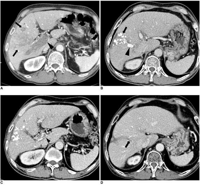
Fig. 1.
Imaging findings are presented for 64-year-old man with hepatocellular carcinoma accompanied by main portal vein invasion.
A. CT scan of portal venous phase obtained at initial presentation shows ill-defined tumor (arrows) in liver parenchyma and thrombus (arrowhead) in main portal vein. Expansion of portal vein and enhancement of thrombus are evident.
B. One month after initial chemoembolization, tumor (arrow) was spotted with iodized oil in liver parenchyma and tumor thrombus (arrowhead) laden along with iodized oil in right anterior portal vein on portal phase.
C. Also noted is decreased extent and diameter of thrombus (arrowhead) in portal vein on same CT scan as B.
D. CT scan of portal venous phase obtained 34 months after last chemoembolization shows small thrombus (arrow) in right anterior portal vein.

