
Figure 4.
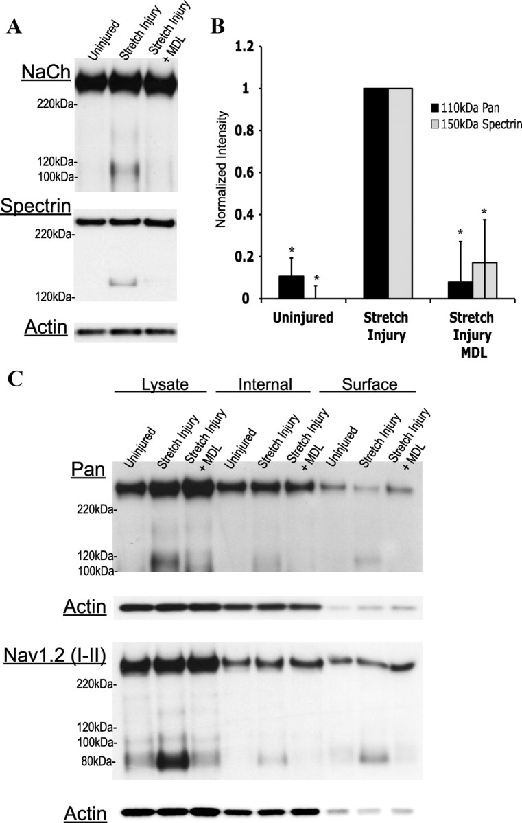
Calpain-dependent NaCh proteolysis occurs in an in vitro model of cortical stretch injury, targeting a specific internal loop of the channel. A, Representative Western blots displaying NaCh proteolysis after a 60–80% stretch injury, with or without pretreatment with the calpain inhibitor (MDL28170; MDL). B, Quantification of the representative blots seen in A. Data normalized to the corresponding stretch injured culture (n = 4, *p < 0.05 from the uninjured control). C, Representative Western blot showing the cellular distribution of proteolyzed NaChs after stretch injury. Six hours after injury, surface membrane biotinylation was performed on cultures with or without a 15 min MDL28170 pretreatment. The 110 and 80 kDa NaCh fragments appeared in the surface fraction, as recognized by the pan and Nav1.2 (I–II loop) antibody, respectively, suggesting that both a C- and an N-terminal portion of the channel remain in the membrane after proteolysis (n = 4).