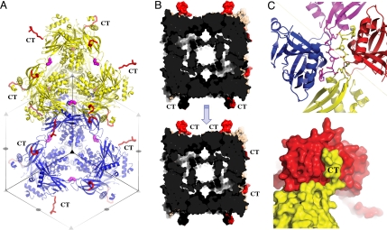
Fig. 4.
Architecture of baculovirus polyhedra. (A) Cartoon representation of two tetrahedral clusters (yellow and blue) showing their tight packing in polyhedra according to the I23 crystallographic translations. Symmetry elements are indicated as gray triangles and ellipses. The C-terminal (CT) loops project outward from the unit cell (red ribbon), and disulfide bonds stabilize the clusters (side chain represented as magenta spheres). (B) Surface representation of two unit cells with the CT loops highlighted in red. These cells pack as indicated by the arrow and interlock with extensive interactions mediated by the CT loop. This forms an extremely dense matrix with only a 30-Å wide cavity delimited by helices H1/2 and no continuous solvent channels. (C) Surface (Bottom) and ribbon (Top) representations of the anchoring interactions mediated by CT loops.

