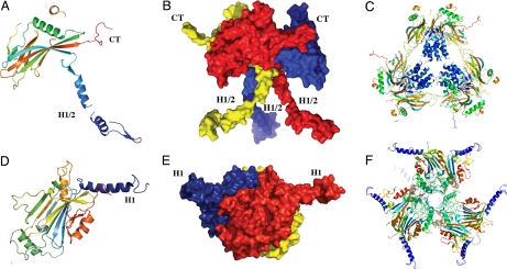
Fig. 5.
Comparison of baculovirus and cypovirus polyhedra. The distinct folds and trimeric organizations of nucleopolyhedrovirus and cypovirus polyhedrin proteins are shown in cartoon (A and D) and surface (B and E) representations. The cartoon representations of the tetrahedral clusters (C and F) highlight the different localizations and functions of the N-terminal helical regions of the two classes of polyhedrins. The central helices H1/2 stabilize the tetrahedral cluster in baculovirus polyhedra, whereas helices H1 extend away from the cluster in cypovirus polyhedra. Molecules are colored in a blue-to-red gradient from the N-terminus to the C-terminus (CT).

