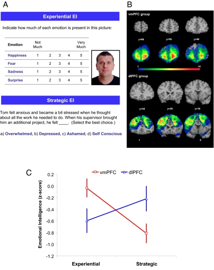
Fig. 1.
Neural substrates of EI. (A) Example items for Experiential and Strategic EI are shown. (B) Coronal views of a healthy adult brain (top and third row), vmPFC group lesion overlap (second row), and dlPFC group lesion overlap (bottom row) are shown in Montreal Neurological Institute space. In each coronal slice, the right hemisphere is on the reader's left. The color indicates the number of individuals with damage to a given voxel. (C) Normalized means (z-scores) and standard errors (SEM) for the Experiential and Strategic EI scores of the vmPFC and dlPFC lesion groups are presented. The individual that appears in A did not participate in the study. The picture is not originally taken from the MSCEIT E-IQ test.

