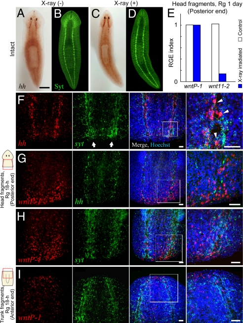
Fig. 5.
Posteriorizing signal originates in differentiated cells (neuron). (A–D) Expression of Djhh was observed in X-ray-resistant differentiated cells of the VNCs. In situ detection of Djhh in nonirradiated (A) and irradiated (C) planarians, followed by labeling of the CNS with anti-DjSynaptotagimin antibody (neural marker; Syt; B and D). (Scale bar, 1 mm.) (E) RGE index of Wnt genes in the posterior blastema of head fragments from X-ray-irradiated animals 1 day after amputation. (F-I) Confocal images of expression patterns of Djhh, DjwntP-1, or Djsyt by dual in situ detection in the posterior (F–H) and anterior (I) end in regenerants from head and trunk fragments, respectively, at 18 h after amputation. Panels show individual expression patterns (red or green color) and their merged image with the image of nuclear staining (Hoechst; blue). The rightmost panel is a magnified view of each boxed area. Arrow indicates VNC (F). Note that Djhh was expressed in neural cells of the VNCs, which were labeled with Djsyt probe (arrowheads; F), but not in the posterior blastema (F and G). Also, DjwntP-1 was predominantly expressed in the posterior end surrounding the VNCs (G and H), but was also weakly expressed in the anterior end (I). (Scale bars, 50 μm.)

