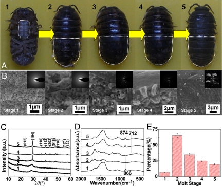
Fig. 1.
Morphology, phase, and composition of cuticles at different molt stages. (A) Photographs of Armadillidium vulgare in the different molt states (1–5). (B) SEM images and SAED patterns of the cross-sections of exocuticle layer shown in A. (C and D) XRD patterns and FT-IR spectra, respectively. (E) Organic contents of the cuticles during the molt process.

