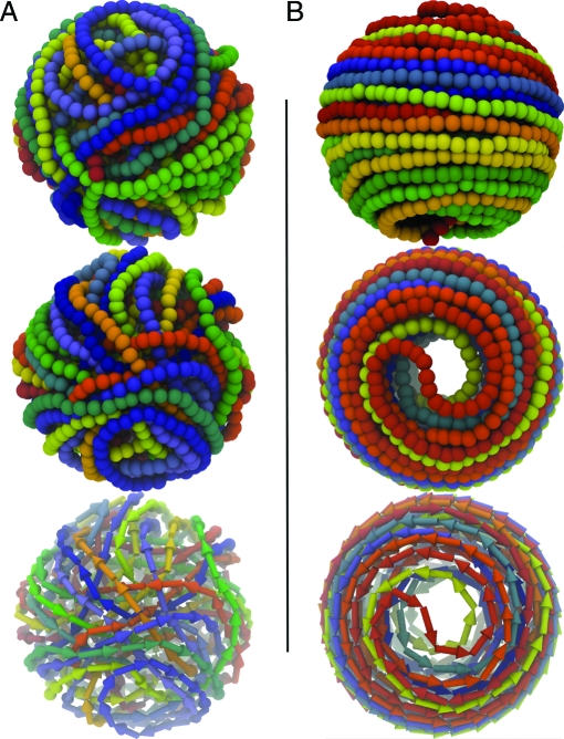
Fig. 2.
Model conformations of the fully packaged 10 kb-long P4 genome in the absence (A) and presence (B) of the cholesteric potential (of strength k c = 1 K B T range Δ = 3 nm and biasing angle α0 = 1°). In each of the two frames, the first two images present front and top views of DNA arrangement. In both cases, a rainbow-coloring scheme (red → yellow → green → blue) is used to follow the indexing of the chain beads (the red end is the one rooted at the portal motor location). The progression of the DNA chain and its winding directonality are also highlighted in the images at bottom, where an oriented arrow is used to represent each triplet of beads.

