
Figure 5.
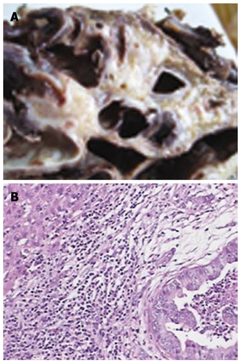
Biliary cystadenocarcinoma. A: Gross appearance of a cross-section of the formalin fixed specimen showing a multilocular mucinous cyst; B: Microscopically, tumor tissues showing moderately differentiated adenocarcinoma with a papillary growth pattern (HE, original magnification × 100).