
Figure 3. Expression of homeostatic chemokines is important for initiation and maintenance of granuloma formation following Mtb infection.
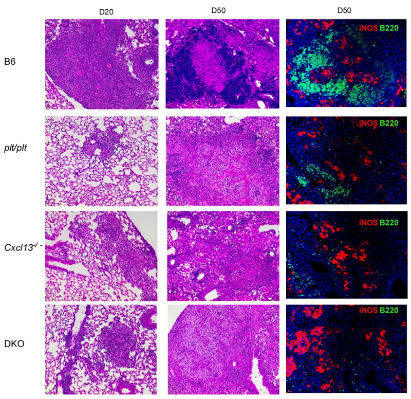
B6 and chemokine deficient mice were infected as described for Figure 1 and lungs were fixed in 10% formalin, embedded in paraffin and stained using H &E as described in Figure 2. The panels in the left column show infected lungs (x10) at day 20 post infection. The panels in the middle column show the cellular components of the granulomata at day 50 post infection (x10). Areas of lymphocyte accumulation appear as darker areas, while areas of macrophage accumulation appear lighter. The panels in the right column show immunofluorescence for B cells (B220 in green) and iNOS (red) on day 50 post infection (x20). One experiment representative of two is shown, n = 4 mice per group.