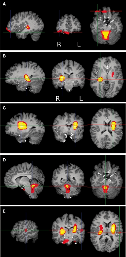
Fig. 1.
Women had relatively larger local volumes in all regions shown except for planum temporale. In axial and coronal images, the left side of the brain is on the right side of the image. (A) Sex differences in the orbitofrontal cortex, coronal view, thresholded at P < 0.001, showing both the right lateral OFC (at crosshair, MNI 34, 40, –5) and ventromedial PFC (center, MNI 6, 36, –12). (B) Sex differences in right WM, near basal ganglia, P < 0.0001, MNI 28, −21, 8. (C) Sex differences in the left basal ganglia and WM, P < 0.0001, MNI −26, −7, 21. (D) Sex differences in the cerebellum, P < 0.0001, MNI 5, −44, −8. (E) Sex differences in the left planum temporale (at crosshair), P < 0.001, MNI −52, −17, 14.

